
CubeLab-Vet
Compact Three-Mode Veterinary Point-of-Care Analyzer
Online ordering is not available yet. Contact sales to confirm inventory, taxes, shipping, and procurement details.
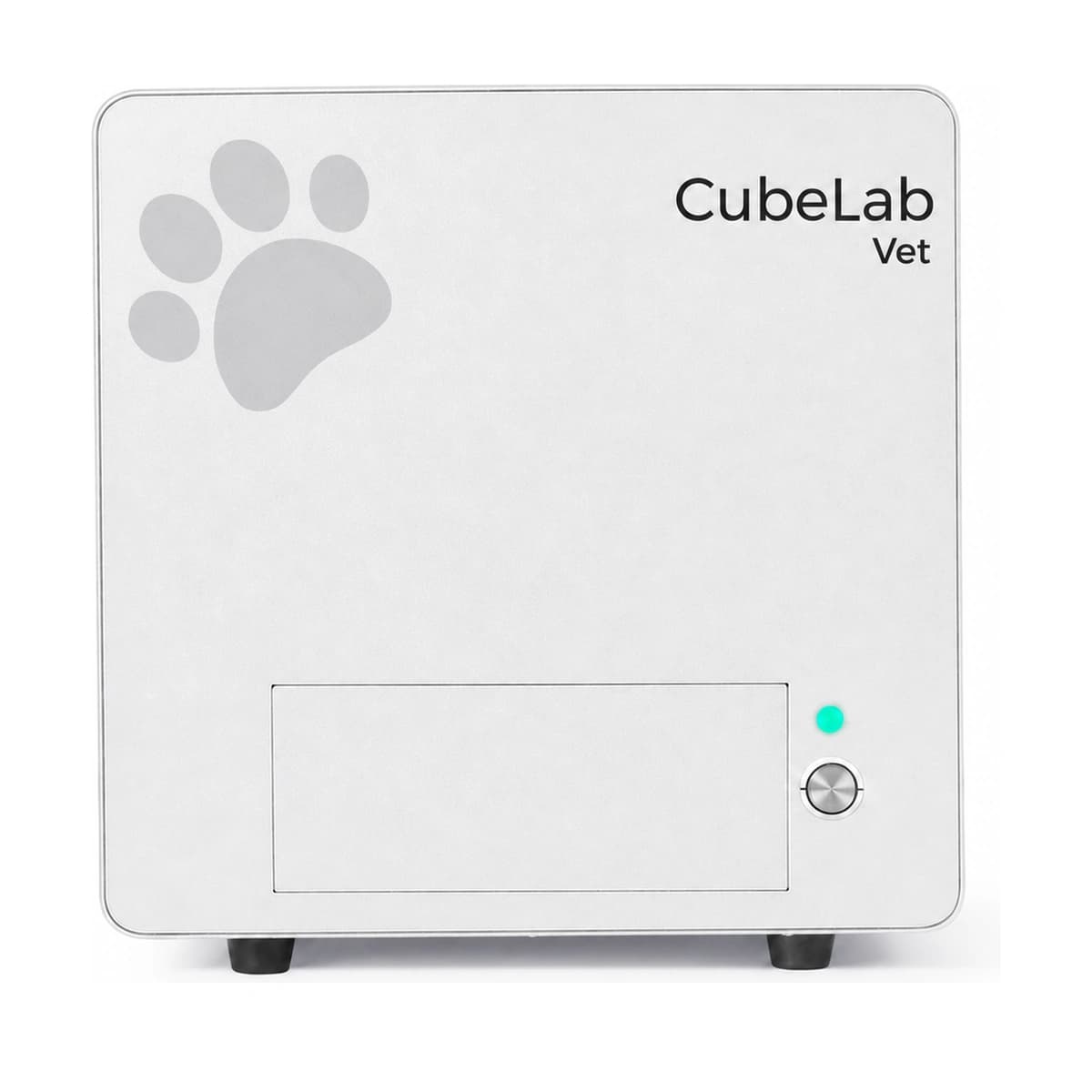
Excl. TaxCubeLab-Vet integrates veterinary biochemistry, fluorescence immunoassay, and dual-mode microscopy within a 33 cm cube. Fluidics-free architecture, single-test lyophilized reagents, automated position and optical calibration, and dual infrared monitoring create a more structured and maintainable veterinary point-of-care workflow.
- Veterinary biochemistry, fluorescence immunoassay, and bright-field/fluorescence microscopy
- No conventional fluidics or wash lines
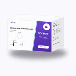
- Single-test lyophilized reagents with 2–30 °C transport and storage
- Automated position and optical calibration
- Dual infrared monitoring and cross-platform App control
* Professional installation and training included.

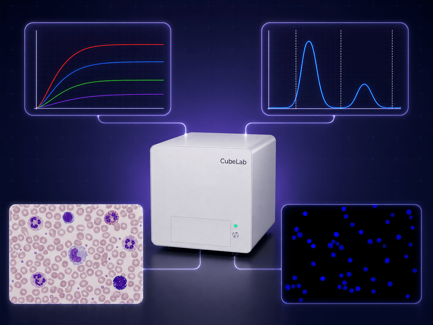
One Instrument. Three Detection Technologies.
Biochemistry, fluorescence immunoassay, and cellular imaging working together in a 33 cm cube
BUILT FOR POINT-OF-CARE TESTING
Bring Laboratory Capabilities into a More Compact Space
CubeLab-Vet combines multi-mode testing, a compact architecture, and software-based control for flexible deployment in veterinary clinics, animal hospitals, and veterinary laboratories.
3
Detection Technologies
Biochemistry, fluorescence immunoassay, and dual-mode microscopy
33 cm
Cubic Footprint
Designed to preserve valuable bench space
18 kg
Instrument Weight
Supports deployment across different testing settings
15–20 min
Combined Multi-Mode Run
Multiple test types within one structured workflow
MULTI-MODE DETECTION
From Soluble Biomarkers to Cellular Morphology on One Platform
Different diagnostic questions require different technical approaches. CubeLab-Vet does not make one method more complicated; it brings complementary detection capabilities into a unified workflow.
Biochemistry and fluorescence immunoassay primarily use serum or plasma. Bright-field and fluorescence microscopy primarily use whole blood. Operators can select the appropriate mode for each assay or combine modes when the workflow requires it.
Biochemistry
Supports common chemistry and related protein assays with a typical time of approximately 10–15 minutes.
Fluorescence Immunoassay
Supports cardiovascular, inflammatory, tumor-marker, and other immunoassay categories in approximately 10–15 minutes.
Bright-Field Microscopy
Uses whole blood for five-part differential CBC testing in under 5 minutes.
Fluorescence Microscopy
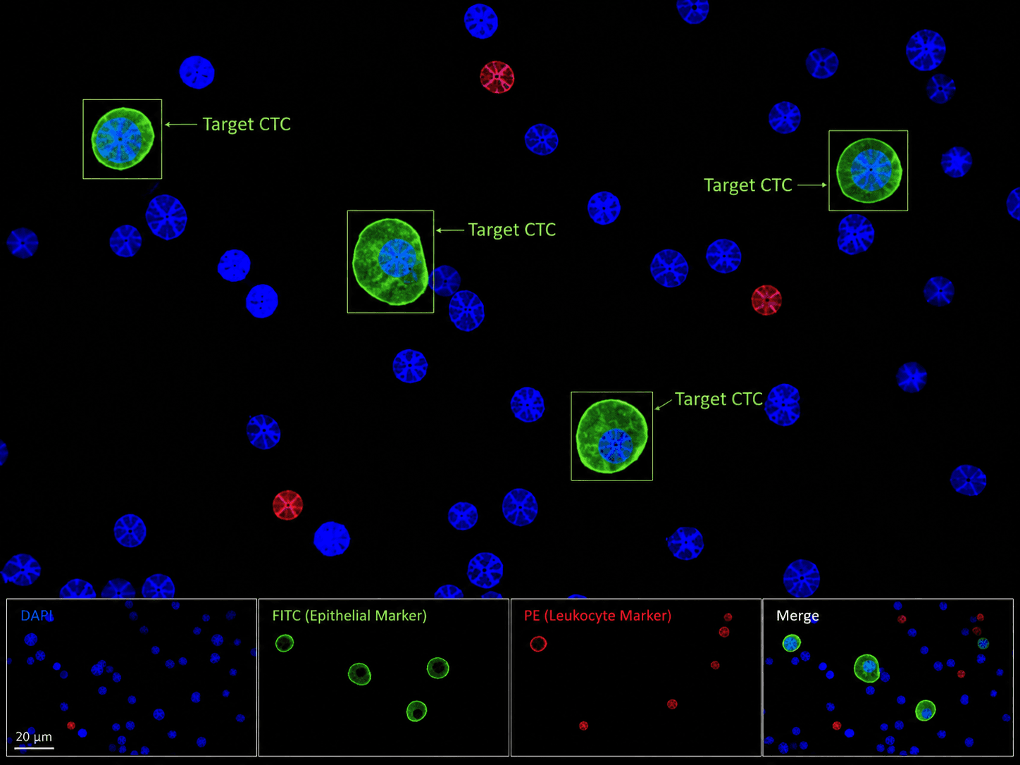
Uses whole blood for fluorescence imaging and assisted identification of circulating tumor cells (CTCs).
BRIGHT-FIELD CELL IMAGING
See Cellular Morphology Behind a Five-Part Differential
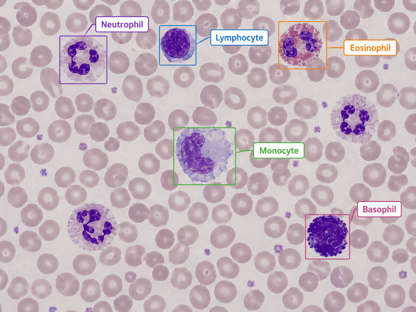
Bright-field microscopy captures cellular images from whole blood and identifies neutrophils, lymphocytes, monocytes, eosinophils, and basophils, providing a visual basis for five-part differential CBC results.
From the original cellular image to the classification output, results remain visually interpretable. CBC testing is completed in under 5 minutes for point-of-care settings that need timely hematology information.
Five-Part Differential
Identifies and differentiates five white blood cell classes to support CBC reporting.
Whole-Blood Sample
A cellular morphology workflow designed around whole-blood specimens.
Under 5 Minutes
Rapid access to hematology information within a compact workflow.
Visual Results
Retains cellular images and classification annotations for review and interpretation.
FLUORESCENCE CELL IMAGING
Multi-Channel Fluorescence Imaging for Assisted CTC Identification
Fluorescence microscopy combines DAPI, FITC, and PE acquisition from whole-blood samples. Merged channels visualize nuclear, epithelial-marker, and leukocyte-marker signals to support identification of circulating tumor cells (CTCs).
Each fluorescence channel reveals a different biological marker. The merged image turns distributed signals into clearer cellular characteristics and supports a structured path from image acquisition and target localization to result review.
DAPI Channel
Visualizes nuclear signals to establish cellular location.
FITC Channel
Visualizes epithelial-marker signals to support target-cell identification.
PE Channel
Visualizes leukocyte-marker signals to help differentiate cell types.
Channel Merge
Combines multiple fluorescence channels for clearer target-cell characteristics.
AUTOMATED PREFLIGHT
Before Testing Begins, the Instrument Checks Itself
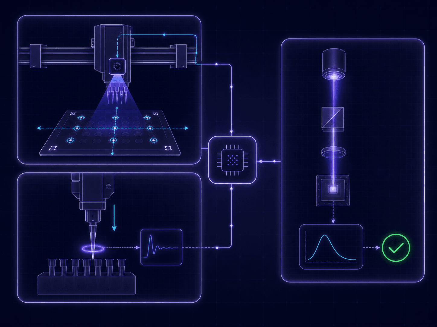
Inspired by the preflight logic of precision automation systems, CubeLab-Vet completes positional calibration, optical calibration, and loading verification before a test begins, reducing preparation steps that depend on operator experience.
01
Position Homing and Calibration
Visual references and motion control confirm key positional relationships and establish a consistent coordinate basis for subsequent operations.
02
Optical State Calibration
The optical modules are checked before testing to establish a stable acquisition baseline for chemistry, fluorescence, and microscopy.
03
Sample and Reagent Verification
Dual infrared cameras help determine whether samples, reagents, and consumables are placed as required.
04
Run When Ready
Testing begins after key conditions are confirmed, with in-chamber monitoring continuing throughout the run.
IN-CHAMBER AWARENESS
Two Infrared Cameras Keep Watch on What Happens Inside
Automation is not only about moving mechanisms. It is about knowing whether each step is happening as expected.
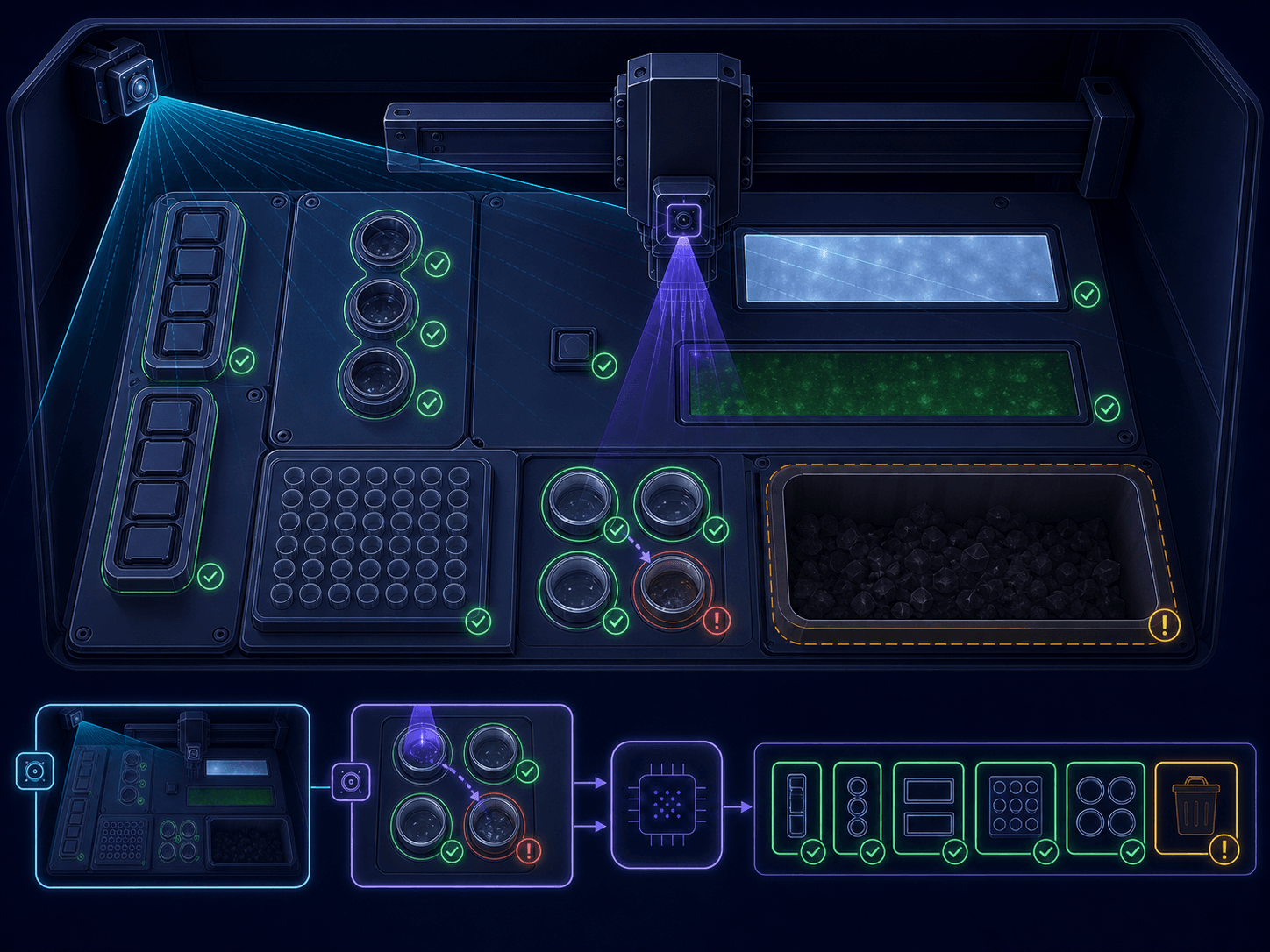
Two infrared cameras cover key operating areas. They verify sample and reagent placement before testing, observe consumable, waste, and abnormal states during operation, and translate conditions that require attention into clear guidance.
Loading Verification
Helps confirm that samples and reagents reach their designated positions.
Consumable Status
Observes consumable preparation and use to reduce missed steps.
Waste Monitoring
Tracks the waste area and provides prompts for subsequent handling.
Exception Awareness
Helps identify in-chamber events that deviate from the expected workflow.
FLUIDICS-FREE ARCHITECTURE
Remove the Fluidics. Remove an Entire Category of Maintenance.
CubeLab-Vet does not depend on conventional fluidics, pumps, valves, or wash lines. A more direct architecture means fewer potential failure points and a clearer maintenance model.
LESS COMPLEXITY
No Conventional Fluidics
Reduces system complexity associated with tubing, pumps, valves, and connectors.
FEWER FAILURE POINTS
Less Risk from Clogs and Leaks
Reduces potential failure points related to clogging, leakage, and aging tubing.
NO WASH LINES
No Wash-Line Dependency
Testing does not rely on conventional wash lines or large volumes of wash solution.
EASIER SERVICE
More Direct Maintenance
Reduces routine work centered on flushing, unclogging, and replacing fluidic components.
SINGLE-TEST LYOPHILIZED REAGENTS
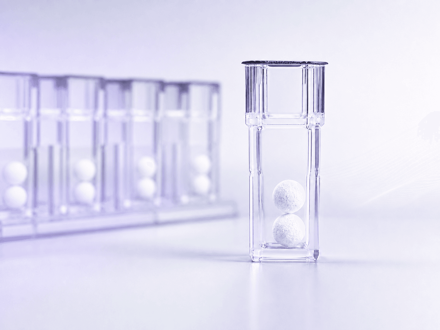
One Independent Lyophilized Reagent for Each Test
The single-test lyophilized format transforms complex liquid reagent management into a clearer, use-on-demand workflow.
Reagents support transport and storage at 2–30 °C, reducing cold-chain dependence. Each test uses an independent reagent, reducing bottle opening, aliquoting, onboard liquid management, and cross-batch handling.
2–30 °C
Ambient transport and storage support deployment across different regions.
Single-Test Format
One reagent unit corresponds to one test for a clear use relationship.
Use on Demand
Reduces bottle opening, reconstitution aliquoting, and liquid reagent management.
POCT Ready
Well suited to distributed, low- to medium-volume point-of-care workflows.
ESIO APP ECOSYSTEM
Control the Instrument from Devices You Already Know
CubeLab-Vet uses the Esio App for connectivity, test control, status review, and result management, keeping the hardware compact while allowing the software experience to continue evolving.
The App supports Windows, iOS, and Android. Depending on the deployment environment, the instrument can connect through Wi-Fi, BLE, USB, or Ethernet for fixed workstations, mobile devices, or local networks.
Cross-Platform
Supports Windows, iOS, and Android.
Multiple Connections
Supports Wi-Fi, BLE, USB, and Ethernet.
Clear Status
Review run stages, instrument status, and operating guidance in one place.
Result Management
Review and manage test results through a unified interface.

COMPACT 33 CM CUBE
Bring Multi-Mode Testing to a Smaller Benchtop
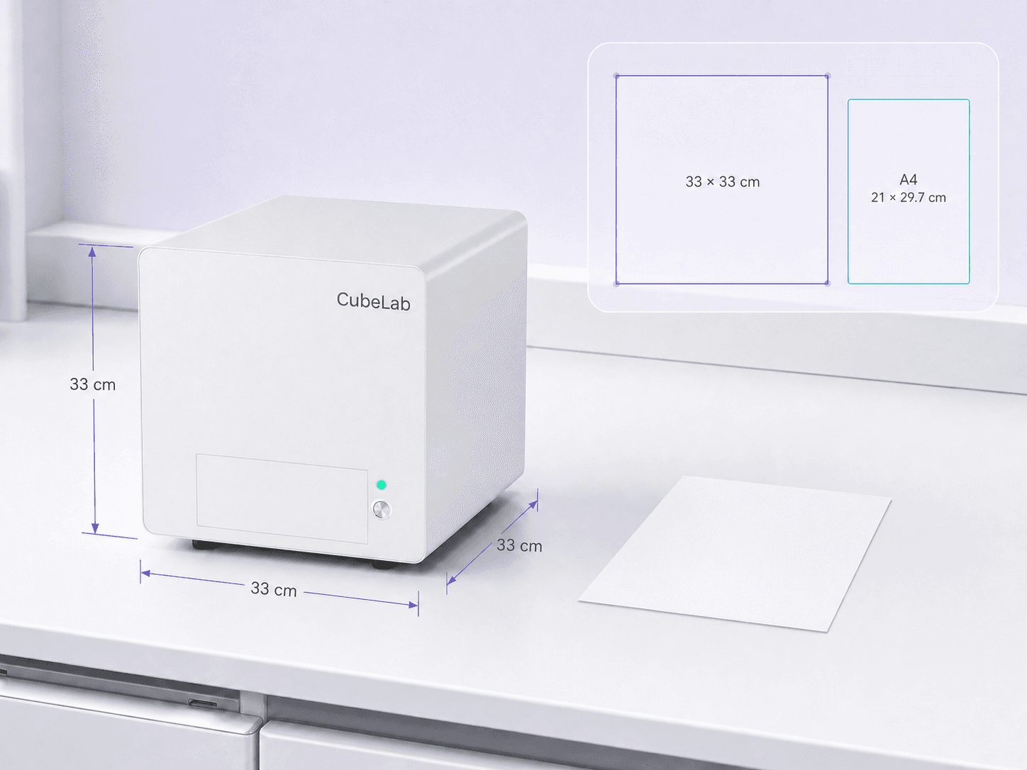
CubeLab-Vet measures 33 cm on every side. Within its compact cubic body, it integrates three detection technologies, automated motion, and in-chamber monitoring while preserving valuable workspace.
A direct comparison with A4 paper makes its footprint easy to understand. The compact form supports deployment in veterinary clinics, animal hospitals, and veterinary laboratories, while allowing work areas to be rearranged as needs evolve.
33 × 33 × 33 cm
A regular cubic form that simplifies benchtop planning.
Approx. 18 kg
Balances structural stability with deployment across different settings.
Compact Footprint
Preserves space for sample preparation and routine operation.
Flexible Placement
Designed for outpatient, primary-care, and smaller-laboratory settings.
POINT-OF-CARE DEPLOYMENT
Bring Testing Capabilities Closer to the Patient
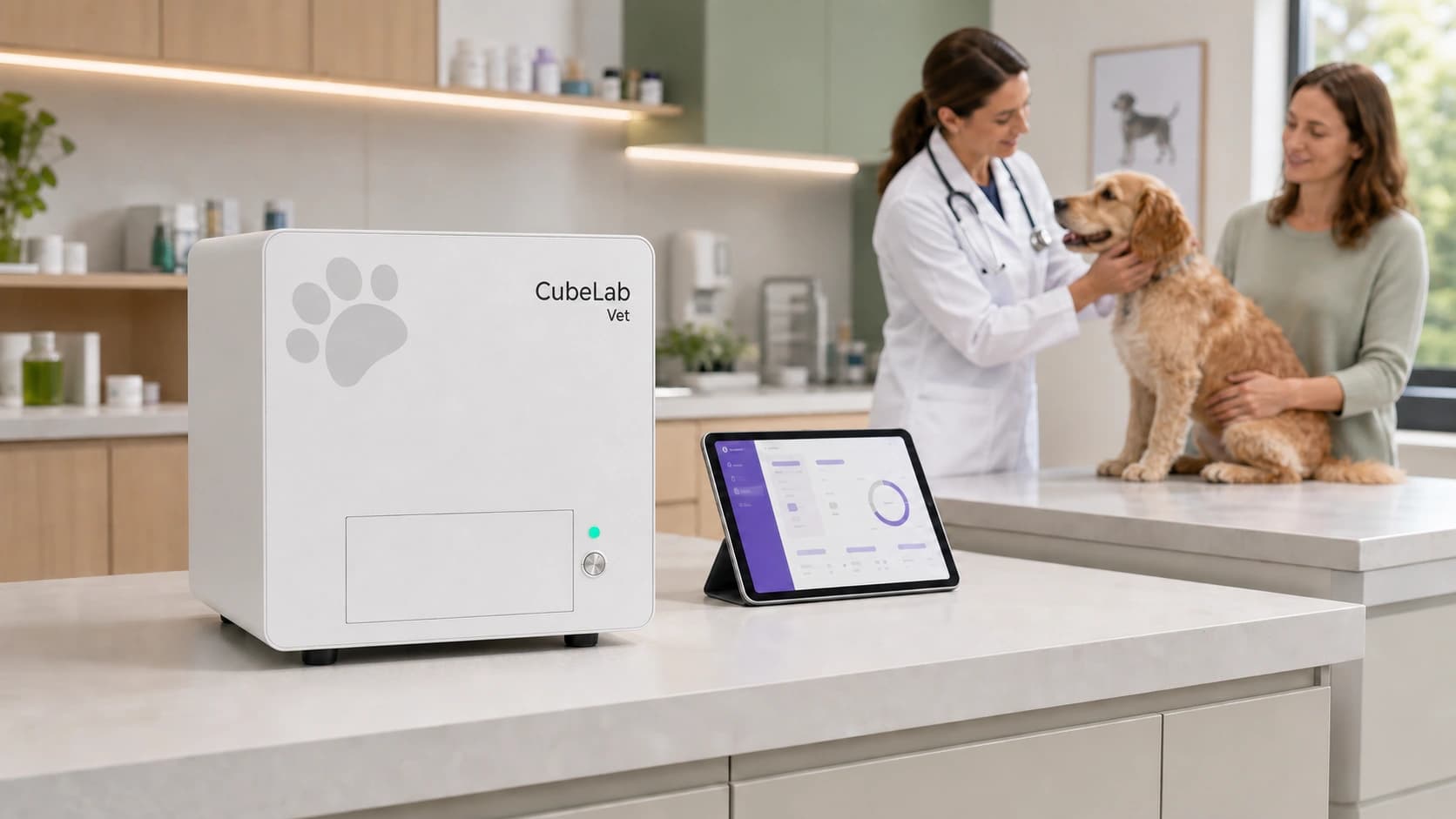
In primary-care and outpatient environments, CubeLab-Vet can be deployed within a conventional clinical workspace, with instrument connectivity, test operation, status review, and result management handled through the Esio App.
Its compact body, fluidics-free architecture, and single-test lyophilized reagents work together to reduce space and maintenance demands, helping multi-mode testing fit more naturally into distributed point-of-care workflows.
Near the Care Setting
Places the testing workflow closer to the patient and clinical team.
App Control
Connects to the instrument and manages the workflow from familiar devices.
No Dedicated Large Room
The compact body fits conventional clinical and small laboratory spaces.
Clearer Maintenance
The fluidics-free architecture reduces work associated with conventional tubing.
| Detection Modes | Veterinary biochemistry, fluorescence immunoassay, bright-field/fluorescence microscopy |
| Primary Sample Types | Serum and plasma; microscopy primarily uses whole blood |
| Biochemistry/Immunoassay Time | Approximately 10–15 minutes |
| CBC Time | Under 5 minutes |
| Concurrent Multi-Mode Run | Approximately 15–20 minutes |
| Reagent Format | Single-test lyophilized reagents; 2–30 °C transport and storage |
| Dimensions | 33 × 33 × 33 cm |
| Weight | 18 kg |
| Control Platforms | Windows, iOS, and Android App |
| Connectivity | Wi-Fi, BLE, USB, Ethernet |